BioEM, Bio-electromagnetism Research Group
CS

Research

Research topics

Basic and applied research on the use of electromagnetic fields in biomedicine for therapeutic and diagnostic applications. Our work also includes the study of interactions of EM fields with living organisms and the determination of safe exposure limits.

  1. Microwave hyperthermia
  2. Non-invasive temperature monitoring during hyperthermia and ablation
  3. Microwave detection and classification of stroke
  4. Measurement of tissue dielectric parameters and phantom development
  5. Radar methods in medicine
  6. Electroporation
  7. Low-field magnetic resonance imaging
  8. Blood-flow assessment and 3D printing of hearts
Therapy · Treatment planning

Microwave hyperthermia
Microwave hyperthermia

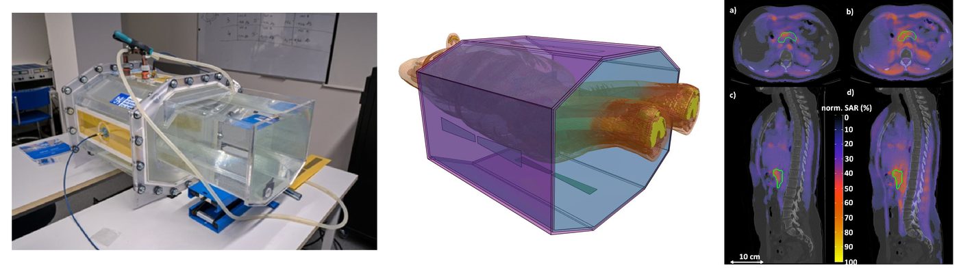
Microwave hyperthermia significantly enhances the effectiveness of radiotherapy and chemotherapy in the treatment of cancer. It allows reduced doses of conventional treatment and thereby minimises side effects.

We are developing a clinical system for microwave hyperthermia focused on the treatment of tumours in the pelvic area, brain, and head and neck. Our work includes:

  • numerical modelling of electromagnetic fields and temperature distribution,
  • development of algorithms for individualised treatment planning,
  • design of applicators, generators and other key components of the system.

Hyperthermia treatment planning tool

We have developed a universal hyperthermia treatment planning tool that enables easy and reproducible creation of simulation models. These consist of 3D patient models created by segmentation of CT series and of a model of the applicator. The tool contains all clinically available regional microwave systems and places the applicator automatically so that the treatment target lies at its centre.

In February 2021 the tool was implemented in the clinical planning workflow of the hyperthermia unit at the Erasmus MC Cancer Institute in Rotterdam and subsequently tested at the LMU university hospital in Munich.

Diagnostics · S-parameters

Non-invasive temperature monitoring during hyperthermia and ablation

Non-invasive temperature monitoring during hyperthermia and ablation

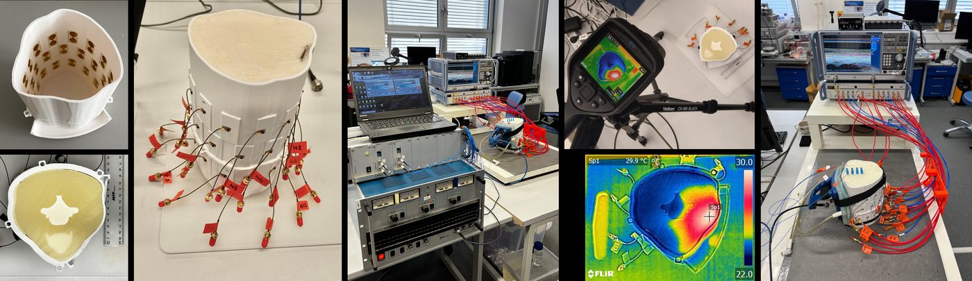
Accurate temperature monitoring during microwave hyperthermia is crucial for the safety and effectiveness of treatment. Invasive measurements provide only point information, while non-invasive methods such as magnetic resonance are financially and technically demanding.

We are developing a system for non-invasive temperature monitoring as part of a comprehensive hyperthermia solution. The research includes:

  • numerical simulations and experimental measurements,
  • reconstruction of the temperature-rise distribution from S-parameters and computed fields,
  • development of models of the temperature dependence of dielectric properties.

We focus on tumours in the pelvis, head, neck and brain, with particular emphasis on hyperthermia of glioblastomas. We therefore intensively study the temperature dependence of the dielectric parameters of brain tissue, which is essential for the accuracy of models and monitoring systems.

Diagnostics · SVM · Born reconstruction

Microwave detection and classification of stroke

Microwave detection and classification of stroke

There is currently no reliable system for pre-hospital detection and differentiation of stroke type (ischaemic vs. haemorrhagic). Rapid diagnosis is crucial for starting the correct treatment and minimising permanent consequences.

Our team is developing a compact, portable microwave system in the form of a helmet that allows:

  • early detection and classification of the stroke type,
  • monitoring of disease progression through differential imaging of changes over time,
  • determining the position and size of the affected brain tissue.

The goal is a device suitable for use in the field, in emergency departments and in ambulances that significantly speeds up decisions about further treatment. For developing your own algorithms we offer the StrokeDataSet from a ten-antenna system.

Metrology · MRI-EPT · Phantoms

Measurement of tissue dielectric parameters and phantom development

Measurement of tissue dielectric parameters and phantom development

Accurate knowledge of tissue dielectric parameters is essential for all our applications, from hyperthermia to microwave-based diagnostics.

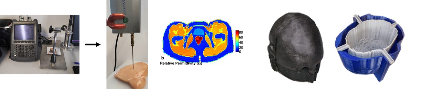
We are developing an affordable measurement system that combines a coaxial probe and a vector network analyser, aiming at rapid evaluation of biological samples, for example immediately after a biopsy. In parallel we create tomographic maps of dielectric properties using the MRI-EPT method. Our activities also include:

  • segmentation of tissues and anatomical structures from CT and MRI data,
  • creation of multi-tissue 3D models of the head, pelvis and whole body,
  • production of realistic biological tissue phantoms with anatomical and dielectric fidelity.

These models and phantoms play a key role in testing and calibrating our therapeutic and diagnostic systems.

Diagnostics · UWB radar

Radar methods in medicine

Radar methods in medicine

We develop radar technologies for medicine, particularly where non-invasive, contactless detection or real-time imaging is crucial.

Radar methods are a promising tool in several clinical and home applications:

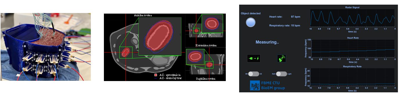
  • Contactless monitoring of vital signs: detection of respiratory rate and heart rate without physical contact, e.g. in intensive care or at home.
  • Navigation systems for catheter insertion: radar imaging assists precise real-time guidance of catheters without ionising radiation.
  • Real-time monitoring of tumour ablation: non-invasive 3D monitoring of the ablation with evaluation of the affected tissue volume directly in the patient.
  • Imaging of metal projectiles (e.g. gunshot wounds): rapid localisation of fragments in the body without CT or X-ray.
  • Fall detection for the elderly: a radar system for automatic fall detection at home and automated assistance alerts.
Therapy · Gene transfection

Electroporation

Electroporation

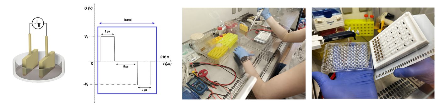
Electroporation, the temporary increase of cell-membrane permeability by an electric field, has a wide range of clinical applications: gene therapy, cancer treatment, targeted drug delivery and biotechnology.

Our activities include:

  • Study of electroporation effects on the target tissue: optimising parameters so that the intervention is both effective and gentle.
  • Analysis of effects on surrounding tissue: monitoring undesirable effects such as haemolysis during cardiac ablation.
  • Numerical simulation of physical phenomena: modelling the electric-field distribution and temperature changes during procedures.
  • Development of hardware and electrodes: new types of electrodes and control units for clinical and laboratory use.
  • Gene transfection: transporting genetic material into cells by electroporation, especially for gene therapy.

The TonaPulse® system developed in the team is commercialised by our spin-off Tonagena, s.r.o.

Imaging · Halbach · B₀ = 50 mT

Low-field magnetic resonance imaging

Low-field magnetic resonance imaging

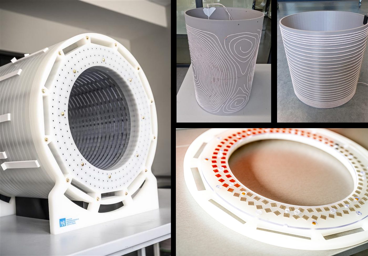
Within the European EURAMET project “Affordable low-field MRI reference system” we collaborate with research institutions on the development of low-cost magnetic resonance systems with B₀ = 50 mT.

The system built at the faculty will enable imaging of the human head and limbs. The magnetic field is generated by a Halbach magnet composed of approximately 2,500 neodymium permanent magnets, so the device will be:

  • significantly smaller and lighter than current clinical MRI scanners,
  • completely passive, and thus with low operating costs,
  • safer for patients with implants.

LF-MRI systems represent a potentially affordable alternative for diagnostics, particularly in resource-limited settings.

Cardiology · CFD · FDM printing

Blood-flow assessment and 3D printing of hearts

Blood-flow assessment and 3D printing of hearts

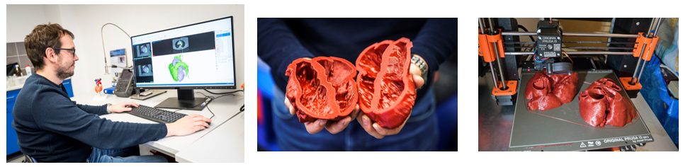
For planning interventional cardiology procedures we segment cardiac structures from CT images and create detailed 3D heart models. In these models we identify up to six possible access points for puncturing the interatrial septum, particularly for closure of the left atrial appendage (LAA), a procedure that plays a key role in reducing stroke risk in patients with atrial fibrillation.

At the same time we perform numerical analysis of flow in the left atrium to explore the impact of LAA morphology on thrombus formation and the likelihood of embolic events. The heart models are:

  • printed with FDM technology from soft plastics,
  • used for planning the optimal procedure,
  • used for simulating blood flow and assessing the risk of thrombus formation around the LAA.